
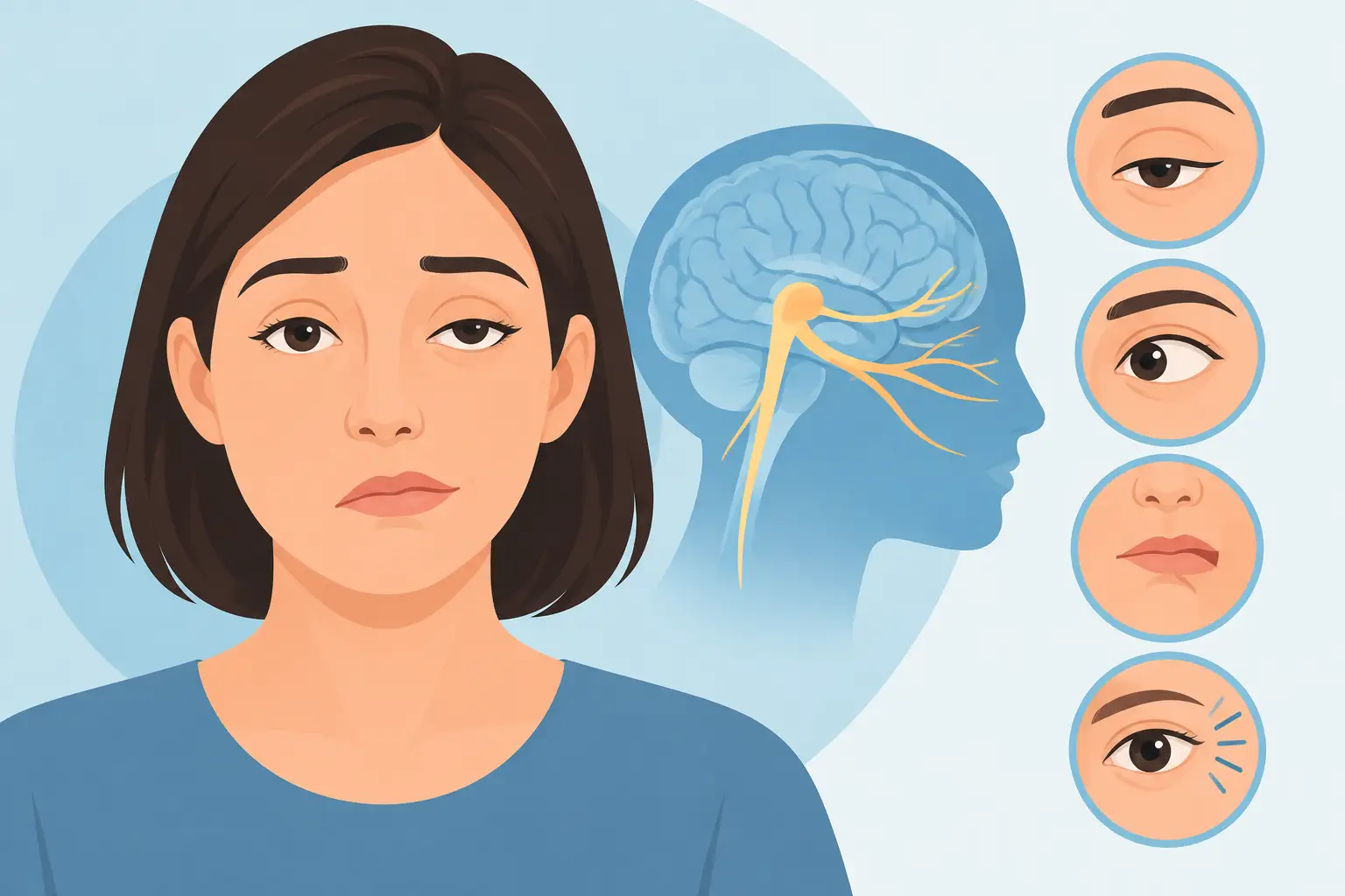
A suddenly drooping eyelid or new double vision is not something to watch for a few days to see if it settles. These symptoms can occur with cranial nerve 3 palsy, a condition that affects eye movement and, in some cases, can signal an urgent neurological or vascular problem.
The third cranial nerve controls several muscles that move the eye, lifts the upper eyelid and helps regulate pupil size. When it is not working normally, the eye, eyelid and pupil may no longer work together as they should. The cause can range from a diabetes-related nerve problem to pressure from an aneurysm, which is why prompt assessment matters.
What does cranial nerve 3 palsy look like?
Cranial nerve 3 palsy is also called oculomotor nerve palsy. It may affect one or more functions of the third cranial nerve, so symptoms vary from person to person. Some people notice a change immediately when looking in the mirror. Others first become aware of double vision when driving, reading or walking down stairs.
A complete palsy often causes the affected eye to sit lower and further out than the other eye. This happens because the muscles controlled by other cranial nerves are still pulling the eye in their usual directions. The upper eyelid may droop significantly, sometimes covering part or all of the pupil.
Common symptoms include a droopy upper eyelid, double vision, difficulty moving one eye up, down or inward, blurred vision, headache or pain around the eye, and a pupil that is larger than the pupil in the other eye. Some people may tilt or turn their head to reduce double vision.
Not everyone will have every symptom. A partial palsy, for example, may cause only subtle double vision or a mild lid droop. Even when symptoms seem minor, a new change in eye movement needs a careful assessment.
When cranial nerve 3 palsy is an emergency
A new third nerve palsy should be assessed urgently, particularly if it begins suddenly. The most concerning combination is a drooping eyelid or double vision with a new, enlarged pupil and severe headache or eye pain. This pattern can occur when an aneurysm puts pressure on the nerve and requires emergency investigation.
Call 000 or attend an emergency department immediately if new double vision or eyelid drooping occurs with any of the following:
- a severe or sudden headache, neck pain, eye pain, nausea or vomiting
- a noticeably enlarged or poorly reactive pupil
- weakness, numbness, facial droop, speech difficulty, confusion or loss of balance
- reduced consciousness, recent significant head injury, or sudden loss of vision
Do not drive yourself if you have double vision, severe pain or neurological symptoms. Ask someone to take you, or call an ambulance if needed.
There is an older clinical rule that a pupil-sparing third nerve palsy in a person with diabetes or high blood pressure may be caused by reduced blood supply to the nerve. While this can be true, it is not a safe reason to delay care. Modern assessment considers the person’s age, symptoms, pupil findings, pain, examination results and risk factors. Imaging may still be needed.
Why does it happen?
The third cranial nerve travels from the brain through the skull and towards the eye socket. Problems anywhere along that pathway can affect it. In adults, reduced blood flow to the nerve is a relatively common cause, especially in people with diabetes, high blood pressure, high cholesterol or vascular disease.
However, clinicians also need to exclude more serious causes, including an aneurysm, stroke, tumour, inflammation, infection, trauma or increased pressure inside the skull. In children, third nerve palsy has a different pattern of possible causes and always requires urgent specialist assessment.
Sometimes no definite cause is found after appropriate testing. This does not make the symptoms less real, and follow-up remains essential. The expected recovery, treatment and need for further testing depend on the suspected cause.
What to expect at an eye assessment
An optometrist or ophthalmologist will begin by asking when the symptoms started and whether they appeared suddenly. Details matter: pain, headache, recent injury, fluctuating symptoms, pupil changes, vascular health conditions and any weakness or speech changes can all guide the next step.
The examination usually includes checking vision in each eye, pupil size and reactions, eyelid position, eye movements and the direction of any double vision. Your practitioner may assess colour vision, visual fields and the back of the eye, and may check blood pressure where appropriate.
If a third nerve palsy is suspected, the priority is determining whether emergency hospital assessment and brain or blood-vessel imaging are required. Depending on the findings, this may involve CT, CT angiography, MRI or MRI angiography organised through hospital or specialist care. Blood tests may also be needed.
At Boda Family Eye Care, a sudden change in eye movement, eyelid position or double vision is treated as an urgent eye concern. An eye examination can identify important signs, but emergency referral is the right next step when symptoms suggest a neurological cause. Bringing a support person can be helpful, especially if your vision is doubled or you are feeling unwell.
Treatment depends on the cause
There is no single treatment for cranial nerve 3 palsy because the treatment is directed at the underlying cause. An aneurysm, for example, may need urgent treatment by a neurosurgical or neurovascular team. A stroke requires immediate hospital management. Inflammatory or infectious causes may need medicines and specialist care.
When the palsy is thought to be related to reduced blood flow in a person with vascular risk factors, management often includes close medical review and addressing diabetes, blood pressure, cholesterol and smoking. Recovery may occur over weeks to months, but the timeline is individual and follow-up is needed to confirm that the nerve is improving as expected.
Double vision can be particularly disruptive during recovery. Covering or blurring one lens of spectacles may provide temporary relief for some people. A prism may be considered once the deviation is suitable and stable enough to measure. For persistent cases, an ophthalmologist may discuss eye muscle surgery or eyelid surgery, but these decisions are generally delayed until recovery has plateaued.
It can be frustrating when the visible eyelid droop improves before eye alignment, or when double vision changes from week to week. Keep follow-up appointments and report any deterioration, new pain, pupil change or neurological symptom promptly.
Practical steps while waiting for care
If you are experiencing double vision, avoid driving, operating machinery or using stairs without care. Closing or covering one eye can reduce the confusion of double images temporarily, but it does not treat the cause. Keep the cover comfortable and alternate sides occasionally if advised by your clinician.
Bring a list of your medications and medical conditions to your appointment or emergency assessment. If possible, note when the symptoms began and whether the eyelid droop, pupil size or double vision has changed. A photo from before symptoms began can sometimes help show what is new, although it should never delay urgent care.
For family members, a new droopy eyelid can be easy to mistake for tiredness or an allergy, especially if there is little pain. If it is accompanied by an eye that does not move normally, double vision, unequal pupils or a headache, seek urgent assessment rather than waiting for a routine eye test.
New double vision or eyelid drooping deserves a clear answer, not guesswork. Prompt examination protects both your vision and your wider health, and gives the right care team the best chance to identify the cause quickly.