
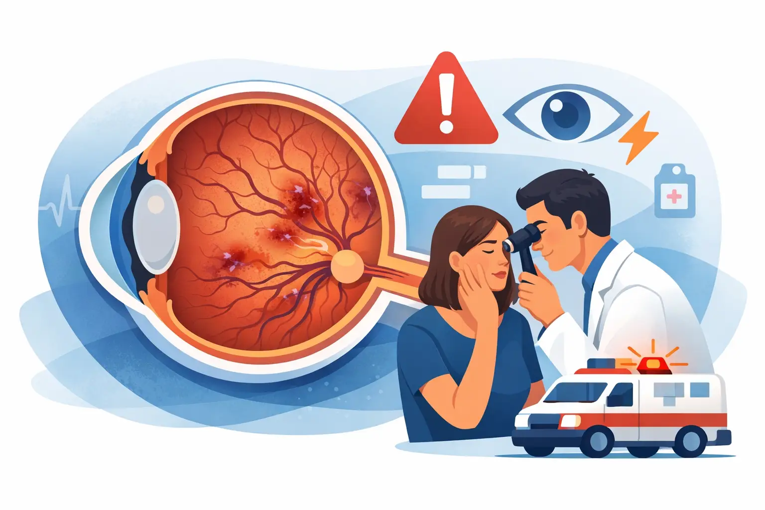
A sudden blur, dark patch or loss of vision in one eye can be frightening, particularly when there is no pain to explain it. Central retinal vein occlusion is one possible cause, and it needs urgent assessment. While vision may change over hours or days rather than in a single moment, waiting to see whether it settles is not the safe option.
At Boda Family Eye Care, we take sudden vision changes seriously. A thorough eye examination can help identify urgent retinal concerns and support prompt referral to an ophthalmologist when specialist treatment is needed.
What is central retinal vein occlusion?
The retina is the light-sensitive layer at the back of the eye. It works much like the film in a camera, sending visual information to the brain through the optic nerve. Blood reaches the retina through arteries and leaves through veins.
A central retinal vein occlusion, often shortened to CRVO, occurs when the main vein draining blood from the retina becomes blocked or compressed. Blood and fluid can then build up in the retina. This may cause bleeding, swelling and reduced oxygen supply to retinal tissue, affecting the clarity and quality of vision.
CRVO usually affects one eye. It is not contagious, and it is not caused by reading, screen use or needing an updated glasses prescription. However, it can be linked with health conditions that affect blood vessels, which is why assessment of both eye and general health matters.
Symptoms that need same-day eye care
Central retinal vein occlusion is often painless. This can make it easy to underestimate, especially if the blur is mild at first. Symptoms vary depending on how much of the retina is affected and whether fluid has collected in the macula, the part of the retina responsible for detailed central vision.
Common symptoms include sudden or worsening blurred vision in one eye, a grey or dark area in the central vision, reduced contrast, distorted straight lines, or a noticeable drop in vision when reading or recognising faces. Some people describe a fog, smudge or shadow that does not clear when they blink.
Any sudden change in vision deserves urgent attention, even if it improves slightly. Similar symptoms can occur with retinal detachment, a tear in the retina, optic nerve problems or other urgent eye conditions. If vision loss is accompanied by facial drooping, weakness, numbness, trouble speaking, severe headache or dizziness, call 000 immediately, as these may be signs of a stroke.
Why does a retinal vein become blocked?
A retinal vein can be affected by changes in the blood vessels, pressure within the eye or factors that make blood more likely to clot. In many cases, there is more than one contributing factor rather than a single clear cause.
The likelihood of CRVO increases with age, but it can occur in younger adults too. High blood pressure, diabetes, high cholesterol, glaucoma, smoking, sleep apnoea and cardiovascular disease can all increase risk. Less commonly, an underlying blood clotting disorder or inflammatory condition may be relevant, particularly for younger patients or those with a personal or family history of clotting.
Having a retinal vein occlusion does not automatically mean a person has had, or will have, a stroke. It can, however, be a useful warning that blood pressure, blood sugar, cholesterol and other vascular risk factors should be reviewed with a GP. Coordinated care between your optometrist, GP and ophthalmologist helps ensure the focus is not only on the eye, but also on longer-term health.
How central retinal vein occlusion is diagnosed
An eye examination begins with listening carefully to what has changed and when. Your optometrist will check vision in each eye, examine the front and back of the eye, assess eye pressure and dilate the pupils where appropriate to obtain a clear view of the retina.
When a retinal vein occlusion is suspected, the back of the eye may show retinal haemorrhages, swollen blood vessels and signs of fluid. Retinal photography and optical coherence tomography, or OCT, can provide detailed images of the retina and help detect macular oedema. This swelling in the macula is a common reason vision becomes blurry.
An optometrist can identify concerning findings and arrange timely referral, but treatment for CRVO is generally managed by an ophthalmologist, often a retinal specialist. The urgency of the referral depends on the examination findings, vision level and whether complications are present.
Treatment focuses on protecting vision
There is no safe home treatment to dissolve a retinal vein blockage. Rubbing the eye, using leftover eye drops or taking unprescribed medicines will not correct the problem and may delay the care you need.
Treatment is directed at complications, particularly macular oedema and the growth of abnormal blood vessels. Many patients are treated with injections into the eye that reduce fluid leakage and swelling. These medicines, commonly called anti-VEGF treatments, may need to be repeated over time. Although the idea of an eye injection can sound daunting, it is performed by an ophthalmologist using numbing drops and careful sterile technique.
In selected situations, steroid treatment may be considered. Laser treatment is not usually used to improve swelling in the central vision, but it can be valuable if abnormal blood vessels develop and threaten complications such as bleeding or raised eye pressure.
The treatment plan depends on the type and severity of the occlusion, the amount of retinal swelling, overall eye health and how the eye responds over time. Regular monitoring is essential, even when vision appears stable, because some complications develop later.
What to do if your vision suddenly changes
If you notice new blur, distortion, a dark area or reduced vision in one eye, contact an optometrist or ophthalmologist for a same-day assessment. Tell the clinic when the symptoms began, whether they are getting worse and whether you have diabetes, high blood pressure, glaucoma or a history of vascular disease.
Avoid driving yourself if your vision is significantly affected. Bring a list of your current medicines and details of your GP or treating specialists if possible. This information can help the care team coordinate any medical review that may be needed.
If you already have a diagnosis of CRVO and develop a further sudden drop in vision, new pain, redness, flashes, floaters or a curtain-like shadow, seek urgent eye care. These symptoms do not always mean the occlusion has worsened, but they should not be ignored.
Can vision recover after CRVO?
Visual recovery varies widely. Some people regain useful vision, particularly when retinal swelling responds well to treatment. Others may have ongoing blur or distortion, especially if there has been significant damage to the macula or reduced blood flow to the retina.
The first few months often provide valuable information about response to treatment, but follow-up may continue for much longer. Keeping blood pressure, diabetes and cholesterol well managed, stopping smoking and attending eye appointments all support better overall vascular and eye health. These measures cannot reverse the original blockage, but they may reduce future risk.
Sudden vision change is never something to put off until your next routine check-up. Prompt assessment gives your eyes the best chance of receiving the right care at the right time.